Part of each umbilical artery closes up degenerating into what are known as the medial umbilical ligaments while the remaining sections are retained as. This fold is formed by the underlying median umbilical ligament.

Mcat Memoranda Umbilical Folds Median Medial And Lateral Are
On this page we have gathered for you the most accurate and comprehensive information that will fully answer the question.
. The median umbilical ligament is a structure in human anatomy. Solutions for Chapter 23 Problem 17WYL. Called also lateral umbilical ligament.
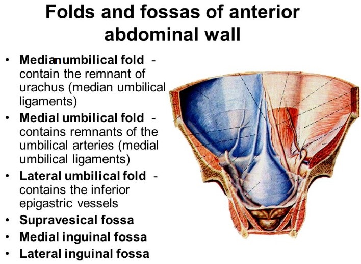
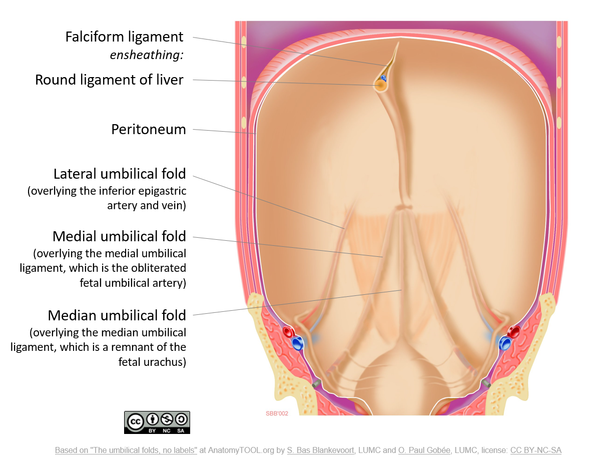
It is also known as the cord of the umbilical artery. The paired medial umbilical folds pass from the pelvis to the umbilicus and cover the underlying medial umbilical ligaments. The medial umbilical ligament is the distal obliterated portion of the umbilical artery.
It is a remnant of the fetal urachus. The median umbilical fold is a raised ridge of parietal peritoneum in the deep aspect of the anterior abdominal wall overlying the median umbilical ligament urachal remnant. It extends from the apex of the bladder to the umbilicus on the deep surface of the anterior abdominal wall.
Each medial umbilical ligament is a remnant of what embryonic vessel. What is the space between the. The allantois regresses into the embryo to give rise to the urinary bladder and median umbilical ligament.
Median umbilical folds. It is one of five umbilical folds and should not be confused with the bilateral. The round ligament which is the remnant of the obliterated umbilical vein runs through the umbilical fissure to connect with the left branch of the portal vein.
The folds are 2 of the 5 umbilical folds and should not be confused with the single midline median umbilical fold. What becomes the median umbilical ligament. It is seen to lie between the transversalis fascia and peritoneum.
It is a fibrous piece of tissue that represents the remnant of the fetal urachus. This duct becomes progressively obliterated during fetal life. It contains the urachus which is an embryonic remnant resulting from involution of the allantoic duct that connects the fetal urinary bladder to the umbilicus.
Solutions for Chapter 23 Problem 17WDL. The medial umbilical ligament is a paired structure found in human anatomy. A fibrous cord sheathed in peritoneum and extending from the pelvis to the navel that is a remnant of part of the umbilical artery in the fetus.
It helps to connect your shin and thigh bones to keep your knee stable and working properly. The median umbilical ligament is a fibrous band located in the anterior portion of the abdomen anterior to the urinary bladder. The MCL medial collateral ligament is a band of tissue that runs along the inner edge of your knee.
Click to see full answer. It is different from the median umbilical ligament a structure that. The median umbilical fold runs superiorly from the apex of the bladder to the umbilicus.
Has two vestigial remnants the ovarian ligament and round ligament which supports the ovaries and uterus in the pelvis. What are the medial umbilical ligament a remnant of. Get solutions Get solutions Get solutions done.
The urachus connects the dome of the bladder to the umbilical cord during fetal life and is located behind. It is on the deep surface of the anterior abdominal wall and is covered by the medial umbilical folds. The medial umbilical ligament is the distal obliterated portion of the umbilical artery.
It develops after birth when the umbilical cord is cut. The median umbilical ligament begins as the allantois in the embryonic period. The medial umbilical ligaments are anatomical remnants of the obliterated foetal umbilical arteries.
It is a shrivelled piece of tissue that represents the remnant of the embryonic urachus. Gubernaculum in the female. The round ligament is sometimes deeply embedded in the umbilical fissure.
Which umbilical fold would bleed if cut. Remnant of umbilical artery. The medial umbilical ligaments are anatomical remnants of the obliterated foetal umbilical arteries.
Where is the medial umbilical ligament. Each medial umbilical ligament is a remnant of what embryonic vessel. This canal is initially open but later closes as the urachus goes on to definitively form the median umbilical ligament.
It extends from the apex of the bladder to the umbilicus on the deep surface of the anterior abdominal wall. The folds are 2 of the 5 umbilical folds and should not be confused with the single midline median umbilical fold. It is a shrivelled piece of tissue that represents the remnant of the embryonic urachus.
Get solutions Get solutions Get solutions done. Just so what are the medial umbilical ligaments remnants of. Thereof what is the medial umbilical fold an embryological remnant of.
What does the lateral umbilical ligament cover. This ligament is also referred to as the cord of the umbilical artery. What is the median umbilical ligament a remnant of.
It then becomes the urachus in the fetus. Lateral to this structure are the medial umbilical ligament and the lateral umbilical ligament. Medical Definition of medial umbilical ligament.
The portion of the vessel gets replaced by fibrous tissue due to the lack of blood flow in the distal part of the umbilical artery. The medial umbilical ligament is an anatomic structure present in the human body that exists as a remnant of blood vessels that were important to fetal circulation. The median umbilical ligament is a structure in human anatomy.
A tubular structure that is a remnant of embryonic development which extends from the umbilicus to the apex of the bladder. An umbilical cord is a thick blood-rich cord that connects a baby to its mother during the gestation process.
![]()
Medial Umbilical Ligament Anatomy Branches Supply Kenhub

Umbilical Folds Wikipedia

Medial Umbilical Ligament Wikipedia

Medial Umbilical Ligament Wikiwand

Internal Abdominal Wall Inguinal Canal Flashcards Quizlet

Positive Med Pg Mnemonics For Remembering Easily Facebook
Umbilical Artery Umbilical Vein 네이버 블로그
The Umbilical Folds And Ligaments English Labels Anatomytool
0 comments
Post a Comment